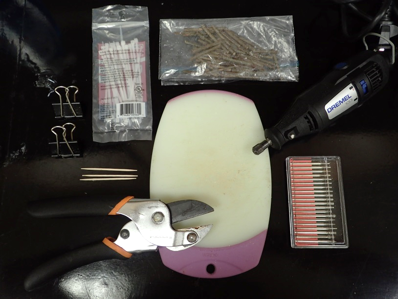
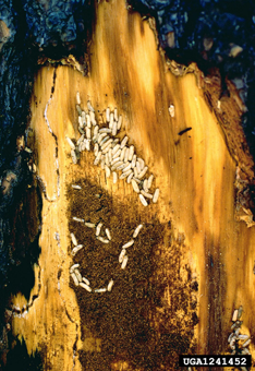
Ambrosia Symbiosis > Lab Protocols
  • Back to Ambrosia Symbiosis Main
  • Lab
  • Create Protocol
  • Edit Protocols
Home» Beetles

Beetles

Wood Flour Media

Posted on April 27, 2018 by Allan Gonzalez in Beetles

Ingredients:
– 75 g of wood flour
– 40 g of agar
– 500 mL of DI water
– 0.35 g of Streptomycin

Instructions:
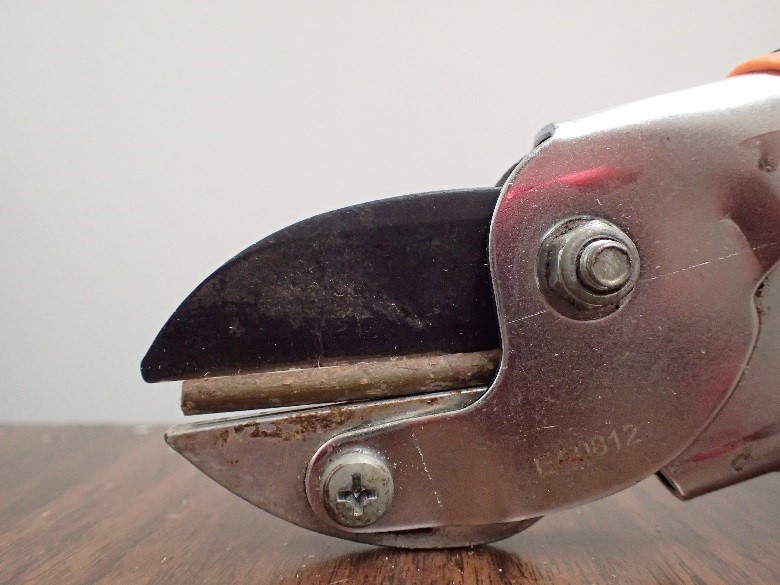
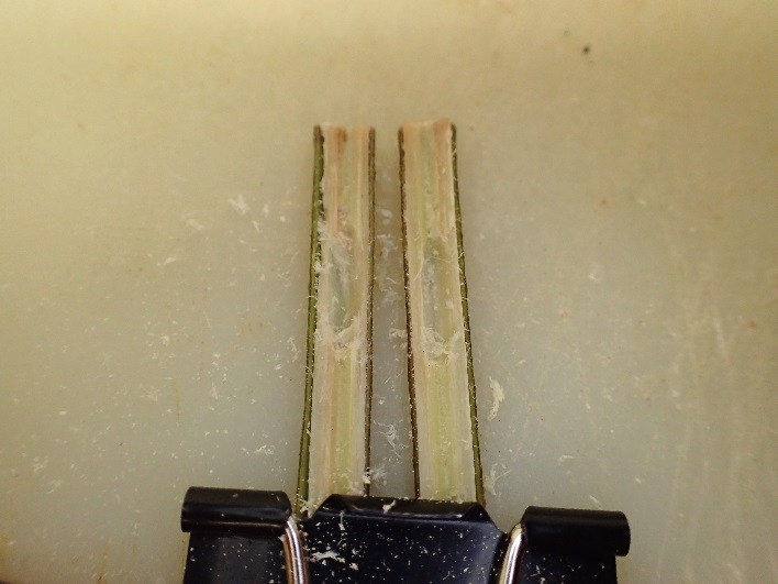
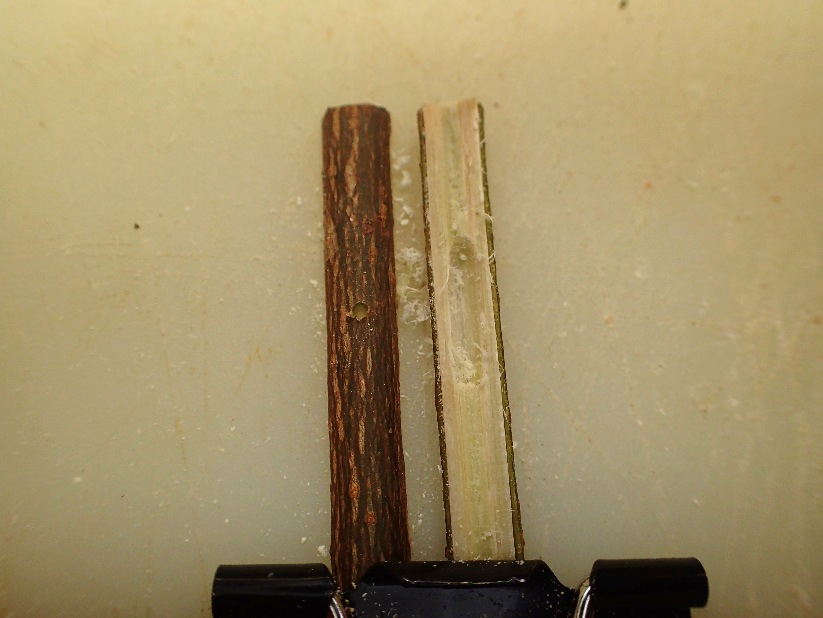
Mix the wood flour, agar, and DI water into an autoclave safe media container. Set the autoclave to 121° C for 30 minutes. Once out of the autoclave, put the container in a hot water bath until it has cooled to 50-55° C. Put the Streptomycin in the media and mix. The media will solidify rapidly so be ready to pour it into plates or tubes once out of the water bath. Scarp the media when it has solidified to make it more habitable to the beetles.

Shipping beetles to the UF Forest Entomology Lab

Posted on April 2, 2018 by jirihulcr in Beetles

Thank you for allowing us to take a look at your beetles! Rest assured that they will be treated professionally.

If you are sending them to be identified for your research, please make sure you provide this information.
If you are depositing the beetles into the UF Forest Entomology collection, here is how they may be used :
– If your samples are of high quality (preserved in high-concentration, non-denatured ethanol from the moment of beetle death, and refrigerated) they will be deposited in -80C and in 99% ethanol, which will preserve them for molecular analyses of the beetles or the fungi vectored by the beetles. They may be used by our team or by anyone else in the global bark beetle research community.
– If your samples are of lesser quality but still morphologically intact, they will be used teaching, such as in the Bark Beetle Academy.

Packaging
1) Please put your specimens in ethanol in a plastic, screw-top vial. Do not use snap-top vials (sometimes called Eppendorf tubes) because they invariably pop open in high altitudes and leak. Do not use glass vials – they break.
2) Add a piece of soft napkin or cotton; this prevents the beetles from trashing around and breaking their legs and antennae.
3) The ethanol is important to dehydrate, clean and preserve the specimens. However it is flammable, so you can’t ship it legally. So after the specimens soak and dehydrate, we recommend pouring off about 90% of the ethanol so you can declare the shipment “dry”.
4) Place your vials on a sheet of paper and attach them with a sticky tape.
5) Put the whole thing into a padded envelope.

Shipping
You can send it cheap regular post. Or feel free to use our Fedex account, so you don’t have to pay anything (happily available on request).
Our lab address is:
Jiri Hulcr
UF School of Forest Resources and Conservation
1745 McCarty Drive
Gainesville, FL 32611
USA
Phone: 517-256-1894

Permits
If you are shipping beetles form overseas, we strongly recommend that you attach a document, confirmed by your institution, stating that the beetles were collected with the appropriate permits issued by the country of origin, and that the sample is provided for mutually beneficial research. (Obtaining permits for dead, dry, common pests should not be difficult.) On the US side, you do NOT need a permit to send such specimens into the us (US regulation 7CFR 330.200).

THANK YOU!

Preparing vials for shipping live beetles

Posted on April 21, 2017 by Zach Nolen in Beetles

Supplies needed:

-2mL Sarstedt screw cap tubes
-Kimwipes
-12.5cm diameter filter paper
-#2 size insect pin
-Flame
-Autoclave

1. For each vial, use pin to poke a small hole in the middle of the bottom from the outside. Do this by heating the pin until red hot on the flame, then pushing it through the center until the tip just breaks into the inside, leaving a small opening between the inside and outside of the tube.

2. Tear off a small piece of kimwipe and push this down to the bottom of each vial.

3. Cut the filter paper into 4 quarters, rolling each one up to about the size of the inside diameter of the tube and push inside. Make sure the filter paper touches the sides and covers everything from the tissue at the bottom to the top of the vial.

4. Cut the excess filter paper off the vial to allow it to close.

5. Fill the tube with loose scraps of kimwipes.

6. After you have a batch of tubes, place them in an autoclave safe container with their caps loose and run on the liquid 15 cycle.

7. If the autoclave wet the tissues inside the tubes, then tightly close them, they are ready to go collecting. If not, add a drop or two of de-ionized water inside each to wet the tissue, then seal tightly.

Artificial Pith Gallery (APG) construction

Posted on July 24, 2016 by Paloma in Beetles

Making Artificial Pith Galleries

Supplies:
– Anvil style pruner
– Small cutting board
– Rotary tool and attachments
– Sections of stem inter-nodes (~4-5cm long, ~0.4-0.5cm thick, sweetgum preferred for X. compactus)
– Cable ties
– Binder clips
– Toothpicks

CH1

1. Collect fresh stem sections, preferably 4-5cm long and 0.4-0.5cm thick.

2. Use pruners to cut stick open, down the middle lengthwise.

CH2

3. Secure each half of stick, cut side up, onto bottom edge of cutting board using binder clips.

4. Use rotary tool with the rounded grinding attachment (See photo) to create a gallery 1-1.5cm in length by removing pith. Make sure not to go past the pith of the wood. Do this on both stick halves so that it is a mirror image.

CH3

5. Use a toothpick to remove any excess sawdust.

6. Take one stick half and flip over, bark side up now, and switch to a fine-point grinding attachment (see photo) to drill the entrance hole for the beetle. This should be done in the center of the gallery.

CH4

7. Remove binder clips and join the two stick halves together, making sure they line up the original way prior to cutting. If done right, each gallery half should come together in the same place to form a full gallery. There shouldn’t be space in the cut between the two halves.

8. Use a cable tie to secure each end of the stick, continuing to hold it together while tightening. Cut excess off with pruner.

9. Freeze until ready to use and then autoclave to sterilize.
CH5

Weekly beetle colony maintenance

Posted on June 23, 2016 by Paloma in Beetles

Daily beetle colony maintenance
I. New females

1. Check black tubes for emergent females. Set aside emergent females. Check collection dates on black tubes. Discard any sticks older than 45 days. Clean emptied tubes with bleach, dry completely, and store in zip lock bags.

2. Thaw frozen sticks for newly emerged females. Seal stick ends with wax (paraffin or parafilm). Place in new sterile petri dishes.

3. Add emergent females, 1 per stick, per petri dish. Label petri dish with date. Place in Starter Box

II. Starter Box

1. Check starter box for excavating beetles. Label dishes with excavating females with the “bored – in” date and unique ID number. Move dish to Working Colony and enter ID and “bored – in” date in spreadsheet.

2. Discard any dishes with dead females, signs of mites, or females that have failed to bore in for longer than 5 days.

III. Working colony

1. Check Working Colony spreadsheet for ripe galleries. Harvest galleries that have reached the “ripe date”. Date will depend on current demand for experiments (ask James). If eggs are needed, this will be 4 – 6 days after “bored – in date”. If pre-pupae are needed, this will be 14 – 16 days after “bored – in” date.

i. Harvesting

1. Place sterile #40 Whitman filter paper in sterile petri dish and moisten with 500 ul sterile PBS.

2. Use sterile pipet tips to transfer eggs/pre-pupae to moistened filter paper, being as fast and sanitary as possible. Add no more than 12 eggs or 6 pre-pupae per filter paper. Wear gloves and clean gloves with EtOH between every stick.

3. If gallery is in good shape and may produce more eggs/pre-pupae, carefully reclose gallery, taking care not to crush beetles. Use small strips of parafilm to bind the ends of the stick back together. Record “opened” date on petri dish and spreadsheet. Return petri dish to working colony. Check 2-3 days later.

4. Discard spent galleries and remove from spreadsheet.

ii. Specimen treatments

1. If current experimental protocol requires axenic larvae/pre-pupae, apply chemical treatment per protocol to eggs/pre-pupae on filter paper (ask James for protocol).

2. Transfer cleaned eggs/pre-pupae to fresh sterile filter paper in new sterile petri dish. Label dish with “harvested” date and chemical treatment, then seal petri dish with parafilm, and move it to the small incubator (27° C).
 
Weekly beetle colony maintenance

Monday: Clean Starter Box and re-supply stocks.

1. Remove all petri dishes

2. Pour perlite into autoclave bag using large funnel and autoclave. Leave
perlite in closed autoclaved bag for use next week.

3. Wipe down container with bleach solution. Dry well.

4. Replace with autoclaved and cooled perlite from previous week.

5. Check and replenish stocks of sterile filter papers, sterile tubes of PBS, media, etc.

Tuesday: Collect fresh galleries.

1. Collect as many occupied X. compactus galleries as possible. Effort will have to be adjusted to match need/loss. Only collect galleries with female visible at entrance.

2. Cut twigs to fit in black tubes. Seal cut twig ends with wax.

3. Place 4-6 galleries per clean black tube. Put tubes in walk-in hot freezer.
Wednesday: Clean Working Colony.

6. Remove all petri dishes/ tubes

7. Pour perlite into autoclave bag and autoclave.

8. Wipe down container with bleach solution. Dry well.

9. Replace autoclaved perlite, petri dishes, and tubes.
Thursday: Collect fresh sticks.

1. Find sweet gum stems 2-6 mm in diameter. Cut sticks ~5 cm length, with no nodes!

2. Collect enough sticks to replace what was used in previous week.

3. Store in zip lock bag in freezer

Collecting Beetles – Red Turpentine Beetles

Posted on May 7, 2014 by caroline in Beetles

For a printer friendly version of these instructions please download the PDF here.

The red turpentine beetle (RTB), Dendroctonus valens, (Figure 1) is a common bark beetle in the U.S. and has recently become a threat to China’s pine forests as an exotic pest. In its native range these beetles are found under the bark of dying or injured pine trees.

Figure 1. The red turpentine beetle.

Known distribution in the U.S.

The RTB is found throughout pine forests in most of the US except for the several states in the southeast (see Figure 2).

Figure 2. Number of recorded RTB collections in the U.S. from T. Atkinson’s curated bark and ambrosia beetle database; http://www.barkbeetles.info/.

Figure 2. Number of recorded RTB collections in the U.S. from T. Atkinson’s curated bark and ambrosia beetle database; http://www.barkbeetles.info/.

Known hosts:

The RTB is ubiquitous wherever pines grow, in any forest, shade, and park trees 8 inches or larger in diameter throughout most of North America (except the South East). Look for resin exudates on freshly cut stumps, the bases of trees that are dying, and trees disturbed by fire, logging, land clearing, or construction.

 Signs of an RTB attack

From a distance, pines with needles that have lost their color and turned red may indicate a potential RTB attack. “The best indicators of red turpentine beetle attack on pines are: large pink-white pitch tubes on the lower bole (Figure 3); accumulations of reddish brown sawdust at the base of the tree and in bark crevices (Figure 4); and accumulations of cream to pink colored crystallized resin granules at the tree base.”

Figure 3. Pitch tubes and resin at the base of a pine from an RTB attack.

Figure 3. Pitch tubes and resin at the base of a pine from an RTB attack.

Figure 4. Sawdust and frass buildup at the base of an RTB attacked pine.

Figure 4. Sawdust and frass buildup at the base of an RTB attacked pine.

Extracting the RTB from wood

RTB  “egg galleries under the bark are fairly wide and linear to irregular in shape. Galleries extend downward from the entry hole 7 cm to 1 m and may even extend into large roots.” Unlike some other bark beetles larval feeding occurs in a gregarious fashion within the gallery.The easiest way to extract RTB from their galleries is to use a wide chisel or the blade of a hatchet to gently lever off  the bark surrounding the pitch tube and entry hole. Once part of the gallery is exposed (Figure 5), beetles can be collected by hand, forceps, or aspirators and deposited in a labeled collection vial. Immediately after collecting, specimens should be stored in high quality ethanol (95%).

Figure 5. RTB adults and feeding larvae inside their gallery under the bark of an RTB infested pine.

Figure 5. RTB adults and feeding larvae inside their gallery under the bark of an RTB infested pine.

Trapping the RTB

Funnel traps baited with a frontalin+turpentine+ethanol lures are the most successful method for trapping the RTB. If needed we can send you a specialized RTB lure from Synergy Semiochemicals. Alternatively, freshly cut pine is a perfect lure as well! If trapping beetles, traps should be checked every day/every-other-day to ensure that fresh, live specimens are being collected for preservation. Immediately after collecting, specimens should be stored in high quality ethanol (95%).

Storing collected beetles

Beetles collected from the same location can stored in the same vial. The vial should could contain 95% ethanol and a label containing collection info. Please store the vial in a freezer until they can be shipped or delivered by another collaborator.

Pictures and quoted text are from the U.S. Forest service Management Guide for Red Turpentine Beetle;  http://www.fs.usda.gov/Internet/FSE_DOCUMENTS/stelprdb5191791.pdf.


Tube Diets

Posted on April 10, 2013 by adamblack in Beetles

Diet composition

Mix:

  • 2.5 g malt extract
  • 44 g sawdust
  • 10 g agar
  • 2.5 g casein (may need to be dissolved in warm water)
  • 5 g dextrose
  • 2.5 g starch
  • Literature recommends adding antibiotics at this step – it makes no difference, since you will autoclave this mixture anyway.

Add 200 ml tap water and mix thoroughly.

Autoclave mixture and pour in skinny long tubes while hot.

Press with something autoclaved, such as the inner part of a syringe, to squeeze out extra liquid.

After pressing media into tubes and letting solidify, have them freeze and defreeze – makes media more aerated.

Notes
Malt extract is pretty acidic, so most bacteria don’t grow there, so antibiotics are not really needed. The bigger problem are molds and mites. Washing beetles can help get rid of some spores, but the beetles come with a rich intestinal microflora and lots of spores anyway, so ultimately the only way to keep your colony from collapsing is starting anew, or re-establishing it with pupae reared on a pure fungus culture.
Mites can be taken care of by keeping your culture on insecticidal paper (available for example from Carolina Biological Company)

Mycelial Diet

Posted on April 10, 2013 by adamblack in Beetles

Protocol

Tanahashi et al. – To obtain the basic artificial diets, agar powder (450 mg) (Nacalai Tesque, Kyoto, Japan), mycelial powder (100 mg), and deionised water containing the preservatives (15 ml) were placed in a 50-ml glass test-tube. Test tubes were autoclaved at 121 °C for 15 min.
Agar with wood only may serve as control.

To obtain mycelia of each fungal species, 5-mm diameter disks of fungal mat were removed from PDA plates, floated singly on 15 ml of aqueous potato dextrose broth (PD broth) (24 g/L) in 50-ml flasks (Sigma Aldrich, St. Louis, MO, USA), and incubated at 25 °C for 10 days. Residues (=mycelia) of fungal suspension by suction filtration were rinsed with deionized water several times and freeze-dried for 24 hours. Dried mycelia were ground with mortar and pestle to 0.5 mm mesh powder and then stored in a desiccator Tanahashi et al., 2009.
Sorbic acid can be used as anti-microbial agent.

  • Sorbic acid (0.828 g)
  • L(+)-ascorbic acid (1.0 g)
  • 133 and sodium hydrocarbonate (1.1 g) in deionized water (1000 ml) (Tanahashi et al., 2009).

Antibacterial additives:
streptomycin sulphate and chlortetracycline hydrochloride, at three different contents of 30, 60, and 300 ppm.Tanahashi et al., 2009 Use no more than 5˚C when using antibiotics.
In Autoclave
Add ascorbic acid to prevent oxygenation of diets in the autoclave. Can add sodium hydrocarbonate to pre-autoclaved diets to raise the pH to ca 5.5.

Extraction from Beetles

Posted on April 10, 2013 by adamblack in Beetles

Protocol

Materials

  • micropipette
  • micropipette tips
  • forceps
  • scalpel
  • razor
  • petri dishes
  • ethanol in petri dish

Procedure

Beetles: 10 female beetles

  1. Remove elytra and clip wings.
  2. Put each beetle in 0.5 ml 1X PBS
  3. Vortex (or sonicate) and keep/plate solution.
  4. In hood, separate beetle abdomen, head, and pronotum
  5. Glue abdomen onto a sterile plate, let dry in hood briefly.
  6. Meanwhile, put head in 0.5 ml 1X PBS
  7. Crush (substitute for extracting mandibular pouches)
  8. Open up the abdomen, suck up the contents under metanotum, transfer it in 0.5 ml 1X PBS’
  9. Remove gut, put in 0.5 ml 1X PBS
  10. and crush.
  11. remove contents from mycangium and transfer in 0.5 ml 1X PBS.

Sonicator test: use six beetles, sonicate two (separately) for 10, 30, 60 seconds. proceed as above. – NO DIFFERENCE, BEETLE STILL LIVING!

Bead beater – fill tubes with 500 μl of PBS. More liquid prevents movement of beads.

Collecting Beetles

Posted on April 10, 2013 by adamblack in Beetles

Collecting tools

The selection of tools partly depends on which types of scolytine beetles you are mostly interested in. For example, for twig bark beetles you will not need any of the heavy duty hardware. On the other hand, trying to pry ambrosia beetles out of a branch with a knife routinely leads to squashed specimens (for xylophages we recommend sawing out a wood “cookie” with the gallery in it, splitting it out with a chisel, and peeling pieces off with clippers, until you get at the beetle). Our recommendations of brands are based on years of experience, not on any relationship with the vendors.

Essential

  1. box cutter (heavy duty)
  2. anvil-style clippers (we recommend Bahco anvil pruner)
  3. hatchet (we recommend Kershaw Camp Axe)
  4. folding saw
  5. broad wood chisel
  6. vials/cryo-tubes with ethanol (screw-top; never use snap-top vials)
  7. pre-cut labels
  8. label pen (we recommend Pigma MICRON archival pen)
  9. soft forceps (we recommend Bioquip Featherweight Forceps Narrow Tip)
  10. tool bag

Not essential but useful

  1. collecting notebook/log
  2. aspirator (pooter)
  3. hard pointed forceps
  4. electric or regular chain saw
  5. scalpel (for very small galleries)
  6. vial box to keep your samples in order

Collection data

levels of humidity

  1. in water
  2. on ground-buried
  3. on ground
  4. above ground- moist
  5. above ground-dry

levels of decay

  1. fresh, sap present
  2. freshly dead, bark easy to peel, sap absent
  3. fungi present, bark loose
  4. bark falls off; other insects
  5. rotten, past main colonization


Bottle trap

Lures
For ethanol lure – attach a mini ziploc bag made of thin plastic, fill half way with ethanol, and poke many hole above the ethanol level.
Take ethanol squirt bottle with you to the field and fill them on site. Change every couple of days (otherwise ethanol will soak up water from the environment).

For dead and well preserved beetles: use upside-down bottle with ethanol in the bottom (no need for ethanol lure bag) here.

For live beetles: use smaller bottle attached to the bottom of flight-intercept bottle. The connector can be made of many things, for example plumbing insulation foam. Cover bottom of the small bottle with shreds of paper towel. Big chunks of paper are not good – then don’t cover the slippery bottom, and beetles have nothing to grab on. Remove live beetles every couple of days (daily is best), and change towel shreds every two weeks at least.

For all beetles: Keep accurate collection information, and keep a card with contact information in case the samples get lost (like a business card). Official UF business cards may be especially important on when travelling by air.
 

Baits

Bait branches
A bait branch bundle is an amazing way to collect twig borers. These are some of the most diverse bark and ambrosia beetles. Make sure you use a meaningful host host. Ideally you would want to pull it up to the canopy, or at least get it off the ground, but not in direct sunlight. The bigger bundle the better! The one on this picture is pretty small.

  • 1
  • 2

View Lab Protocols

  • expandBacteria
  • expandBeetles
  • expandCurrent Work
  • expandDatabase
  • expandFungi
  • expandGeneral Molecular Work
  • expandMiscellaneous
  • expandPathology
  • expandSequence Analysis

(c) 2013 Ambrosia Symbiosis Lab Protocols

Cleantalk Pixel